Summary
A bone scan (skeletal scintigraphy) is an imaging modality in which an intravenously administered radioactive tracer with an affinity for bone (e.g., Tc99m-methylene biphosphonate) is used to visualize areas of increased metabolic activity within bone with the aid of a gamma camera. Bone scans are used to evaluate bone injuries, musculoskeletal pain, joint conditions, primary bone neoplasms, and malignancies with potential for bone metastases.
Indications
A bone scan is used for the detection, evaluation, and/or follow-up of the following conditions:
- Primary malignant bone tumors (e.g., osteosarcoma)
- Bone metastasis from primary extraosseous tumors (e.g., breast cancer, prostate cancer, lung cancer)
- Inflammatory and/or degenerative disorders; (e.g., osteomyelitis, rheumatoid arthritis, psoriatic arthritis)
- Paget disease of bone
- Fractures (e.g., stress fractures, nonaccidental injury in children, fracture healing)
- Impaired vitality of bone (e.g., infarct of bone transplants, osteonecrosis)
- Bone diseases that do not show up on primary imaging studies (e.g., x-ray, CT)
References:[1]
Contraindications
- Absolute contraindication: pregnancy
-
Relative contraindications
- Breastfeeding women
- Pathology with a predominantly osteoclastic process (e.g., multiple myeloma)
- Trauma or surgery in the region of interest within the past 6–12 months .
References:[2]
We list the most important contraindications. The selection is not exhaustive.
Technique/steps
- Intravenous administration of a radionucleotide tracer with an affinity for bone (e.g., Technetium-99m diphosphonate, Technetium-99m oxidronate)
- Uptake of tracer in areas with high bony metabolic activity (“hot spots”)
- Uptake of the radiotracer by a particular region of the bone is dependent on:
- Regional blood flow
- Osteoblastic activity
- Amount of osteoid
- Uptake of the radiotracer by a particular region of the bone is dependent on:
- Detection of these “hot spots” with a gamma camera
- Renal elimination of the radiopharmaceutical agent with increased uptake in the urinary tract
References:[2][3]
Interpretation/findings
-
Very high sensitivity for the detection of areas of bone with increased metabolism
- The following conditions might not be visualized on a bone scan:
- Tumors confined to the bone marrow
- Rapidly growing tumors with slowly regenerating bone (e.g., plasmacytoma)
- Osteolytic metastases
- The following conditions might not be visualized on a bone scan:
- Low specificity


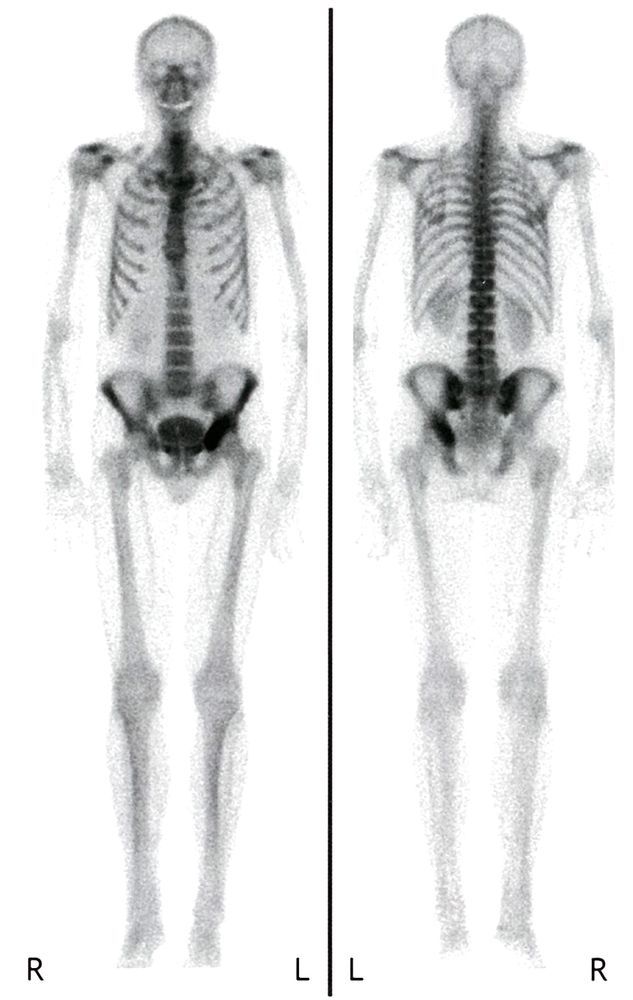
Bone scan (Tc-99m-MDP, whole body) of a patient with a history of chondrosarcoma of the pelvis
Intense uptake of radiopharmaceutical (indicated by black arrowheads) is visible in the region of the mass in the left ischium.
Chondrosarcomas frequently display intense uptake on bone scintigraphy.
Source: © IMPP
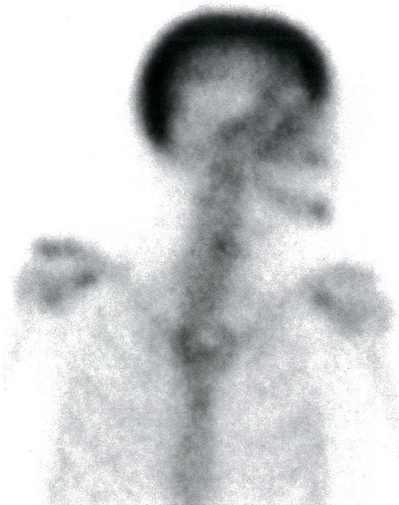
Nuclear medicine bone scan upper body (technetium 99m-methyl diphosphonate (Tc99m-MDP); anterior view) of a patient with Paget disease of bone (osteitis deformans)
Intense radionuclide uptake is seen in the calvarium (red overlay).
Tc99m-MDP bone scans are sensitive, but not specific, for Paget disease. Increased radionuclide uptake may be seen during all phases of the disease, especially when a lesion is active; uptake may be normal during the quiescent osteosclerotic phase. Bone scans are sensitive for the detection of increased blood flow and osteoblastic activity, and are very useful in defining the overall extent and distribution of Paget disease.
Source: © IMPP
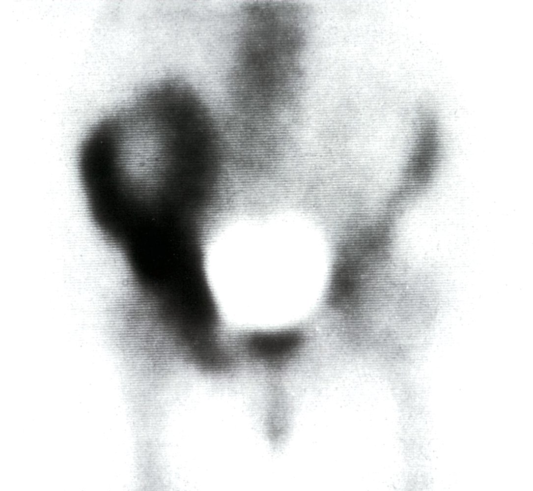
Nuclear medicine bone scan pelvis (technetium 99m-methyl diphosphonate (Tc99m-MDP); anterior view) of a patient with Paget disease of the bone (osteitis deformans)
Intense radionuclide uptake is seen in the right hemipelvis (red overlay).
Tc99m-MDP bone scans are sensitive, but not specific, for Paget disease of the bone. Increased radionuclide uptake may be seen during all phases of the disease, especially when a lesion is active; uptake may be normal during the quiescent osteosclerotic phase. Bone scans are sensitive for increased blood flow and osteoblastic activity, and they are very useful in determining the overall extent and distribution of Paget disease.
Source: © IMPP
References
- "ACR-SPR practice parameter for the performance of skeletal scintigraphy (bone scan)". https://www.acr.org/-/media/ACR/Files/Practice-Parameters/Skeletal-Scint.pdf. [2017-01-01]
- "Clinical guideline for bone scintigraphy". https://www.bnms.org.uk/images/stories/Procedures_and_Guidelines/BNMS_Bone_Scintigraphy_Guideline_v1.pdf. [2018-10-01]
- Love C, Din AS, Tomas MB, Kalapparambath TP, Palestro CJ. "Radionuclide bone imaging: an illustrative review". Radiographics. 23(2). :341-358. (2003)