Summary
Hypovolemia refers to a state of intravascular volume depletion, while dehydration describes a state of reduced total body water volume, mostly affecting the intracellular fluid compartment. In clinical practice, however, these terms are often used interchangeably, as they are often encountered simultaneously. Body fluid loss (dehydration and/or hypovolemia) occurs when fluid excretion exceeds fluid intake, e.g., due to inadequate fluid intake, vomiting, and/or diarrhea. Young children and the elderly are at an increased risk of clinical dehydration because of differences in body water distribution, the potential inability to communicate needs to caregivers, and increased diuretic use in elderly patients. Patients may present with increased thirst, lethargy, prolonged capillary refill, abnormal vital signs, and decreased skin turgor. Patients can also develop hypovolemic shock if hypovolemia is so severe that the body is unable to compensate, resulting in end-organ damage due to hypoperfusion. Hypovolemia and dehydration are clinical diagnoses and laboratory tests are only indicated in patients with suspected associated metabolic disturbances or severe enough fluid loss to cause end-organ damage. The primary goals of treatment are to first address the hypovolemia, if present, in order to quickly restore the circulatory volume, followed by the management of dehydration through the gradual correction of any remaining fluid deficit (including free water deficit), associated electrolyte abnormalities, ongoing fluid losses, and maintenance fluid requirements.
See also “Intravenous fluid therapy” and “Shock.”
Overview
Dehydration and hypovolemia often occur together, however, there are significant pathophysiological and clinical differences between the processes that can affect management.
| Overview of dehydration and hypovolemia[1][2][3] | ||
|---|---|---|
| Dehydration | Hypovolemia (extracellular volume depletion) | |
| Typical causes[1][4] |
|
|
| Fluid loss |
|
|
| Compensatory mechanism |
|
|
| Clinical features |
|
|
| Diagnostics (See “Laboratory findings in hypovolemia and dehydration” for details) |
|
|
| Treatment |
|
|
Etiology
- Insufficient water intake, particularly in elderly individuals and the critically ill
-
Increased free water loss
- Renal loss: diuretics, hyperglycemia, polyuric phase of renal failure, diabetes insipidus
- Extrarenal loss: diarrhea , vomiting, burns, increased sweating, fever, inflammation, ascites
- See also hypovolemic hypernatremia and euvolemic hypernatremia.
References:[5][6][7][8][9]
Clinical features
Common features
- General symptoms include thirst, headache, weakness, dizziness, and fatigue.
- Physical findings often include:
- All patients: dry skin, dry mucus membranes, and reduced tears
- Infants: sunken fontanelle, irritability, and lethargy
- Hemodynamic instability may be present in severe cases
Estimating severity
- Dehydration and hypovolemia often coexist in the same patient (see “Overview” for a comparison of clinical features and etiology).
- Estimating overall fluid loss (due to both processes) is helpful to stratify clinical severity upon presentation
- Estimate percent weight loss based on clinical features.
- For children ≤ 5 years of age with gastroenteritis, consider using the clinical dehydration scale (see “Infectious gastroenteritis in children”).
- Severe fluid loss is a cause of hypovolemic shock (see also “Clinical features of shock”).
| Clinical features of dehydration and hypovolemia[1][10] | ||||
|---|---|---|---|---|
| Clinical features | Mild fluid loss (3–5% weight loss) | Moderate fluid loss (6–9% weight loss) | Severe fluid loss, i.e, hypovolemic shock (≥ 10%weight loss) | |
| Symptoms | Behavior and activity level |
|
|
|
| Thirst |
|
|
|
|
| Physical findings | Vitals |
|
|
|
| Eyes |
|
|
|
|
| Skin |
|
|
|
|
| Mucous membranes |
|
|
|
|
| Urine output |
|
|
|
|
| Anterior fontanelle (infants only) |
|
|
|
|
| ||||



10-year old boy
The acute presentation of sunken eyes, especially in association with altered mental status or unconsciousness, should raise suspicion for potentially life-threatening dehydration.
The cause for dehydration in this child was diarrhea due to typhoid fever.
© AMBOSS
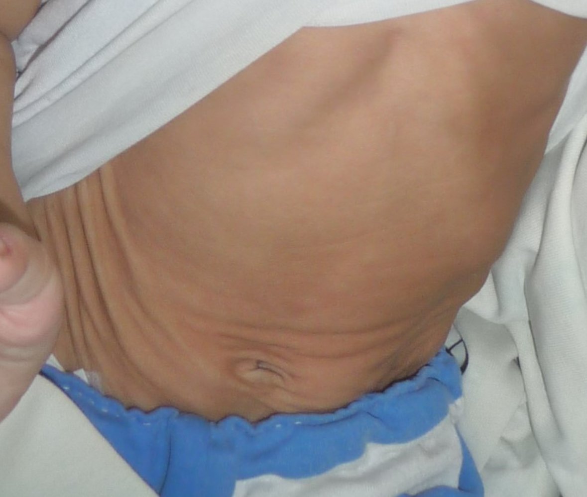
Abdomen of an infant: After pinching, the abdominal skin recoils slowly, showing significant skin tenting. This may be a sign of severe dehydration, e.g., in the course of a gastrointestinal infection.
© AMBOSS
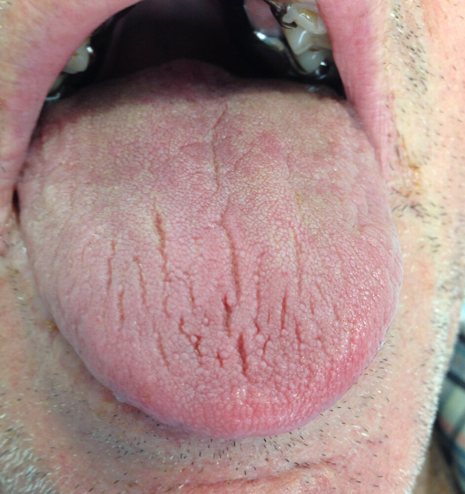
Photograph of a patient with xerostomia
The tongue of the patient shows several longitudinal furrows of different depths. This finding is typically caused by dehydration.
Source: “Xerostomía” by Pilar Molés Julio, Wikimedia commons, licensed under CC BY-SA 4.0.

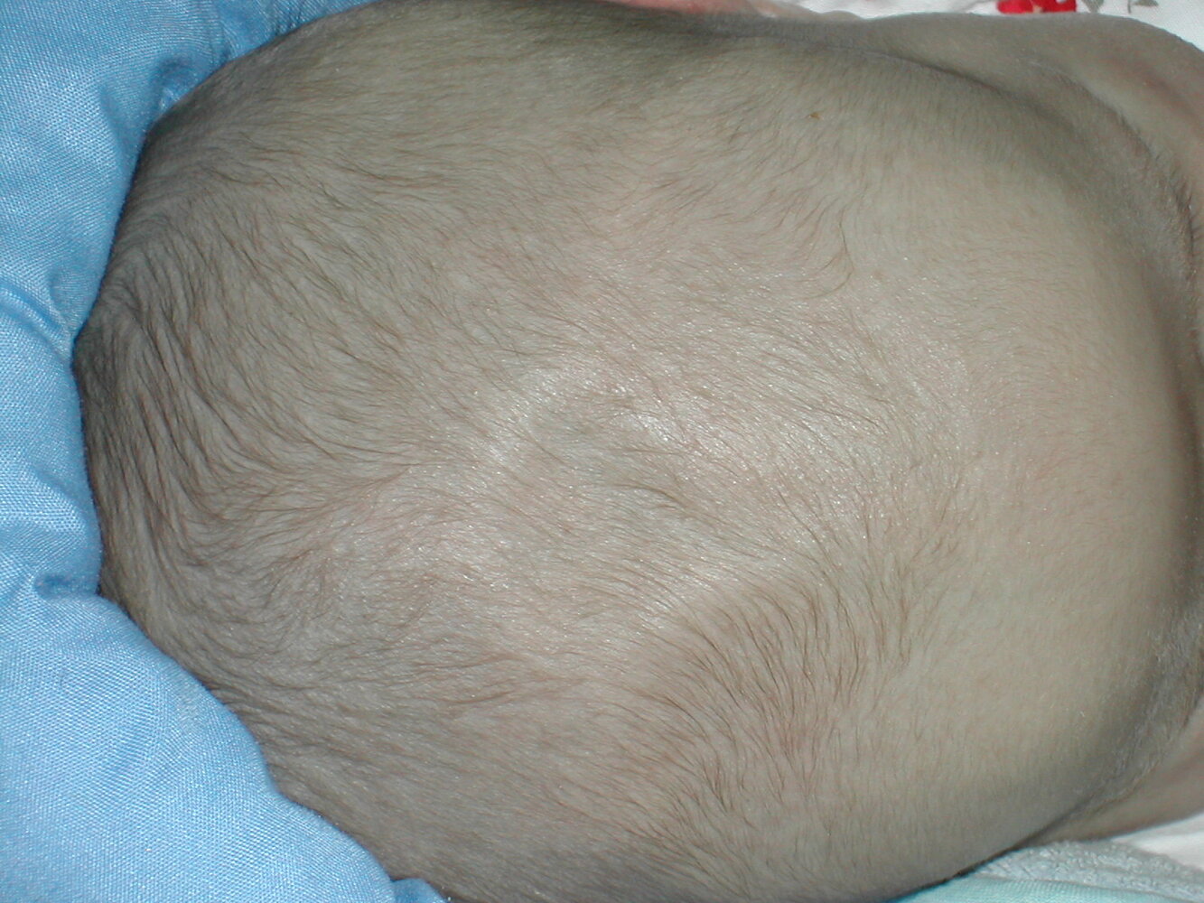
Head of a 1-month-old infant
The skin overlying the anterior fontanelle is notably indented. This finding typically indicates dehydration, especially if other signs of dehydration, such as decreased wet diapers and poor feeding, are present.
Source: “Human anterior fontanelle 1 month dscn1449” by Johann Dréo, Wikimedia Commons, licensed under CC BY-SA 2.0.

Diagnosis
Approach
- Diagnosing dehydration and/or hypovolemia relies on clinical assessment (see “Clinical features of dehydration and hypovolemia”).
- Laboratory studies can help support the clinical diagnosis but are not routinely required.
- Obtain laboratory studies for:
- Severe fluid loss requiring IV fluids
- Moderate fluid loss following unsuccessful enteral (e.g., PO/NG) fluid replacement
- Evaluation of the underlying cause, e.g., diabetes insipidus, hyperglycemic crises
Dehydration and hypovolemia are clinical diagnoses.
Laboratory studies
-
Orders: to evaluate severe fluid loss
- BMP: Serum Na used to guide treatment.
- CBC
- Lactate
- Uric acid
- POC glucose
- Urinalysis
- Urine electrolytes: e.g., urine Na
-
Findings
- Interpret in context on an individual basis
- Vary depending on: [2]
- Predominance of dehydration vs. hypovolemia
- Age: e.g., infants, older adults
- Underlying etiology: e.g., diabetes insipidus
- Medications: e.g., diuretic use
- Comorbidities: e.g., CKD
- See also “Diagnostic findings in hypernatremia.”
| Laboratory findings in dehydration and hypovolemia[2] | ||
|---|---|---|
| Dehydration | Hypovolemia | |
| Plasma |
|
|
| Urine |
|
|
Treatment
Replacing body fluid losses typically involves rapid correction of extracellular volume depletion and judicious correction of intracellular dehydration (see also “IV fluid therapy strategies”).
Approach
-
Initial fluids for dehydration and hypovolemia: Administer isotonic fluid and choose route and rate based on estimated fluid loss.
- Severe fluid loss; (hypovolemic shock): Begin immediate hemodynamic support with aggressive IV fluid resuscitation, e.g., 20 mL/kg bolus of isotonic crystalloid.
-
Moderate fluid loss (hypovolemia without shock)
- Administer enteral OR parenteral fluids to correct extracellular volume deficit. [11]
- If starting with IV fluids, transition to enteral fluids as soon as possible.
- Mild fluid loss: Prioritize enteral replacement of fluids (i.e., oral rehydration therapy).
-
Supportive care for all patients: Management of these is often begun concurrently with initial fluid administration.
- Treat associated metabolic disturbances: e.g., glucose and electrolyte abnormalities.
- Identify and treat underlying causes (see “Etiology”).
- Address continued fluid needs.
- Replace free water deficit to restore intracellular volume losses and correct hypernatremia.
- Meet maintenance fluid requirements.
- Replace ongoing fluid losses.
-
Monitoring and disposition
- Evaluate the need for hospital admission (see “Disposition”).
- Inpatient monitoring
- Regular vital sign monitoring
- Regular input/output monitoring
- Consider serial serum electrolytes.
Stabilization through correction of intravascular volume deficit with fluid resuscitation is the first priority. Manage urgent metabolic abnormalities (e.g., severe symptomatic hyponatremia, acute hypoglycemia) concurrently with fluid resuscitation. Address subacute electrolyte abnormalities after stabilization.
Initial fluid therapy for dehydration and hypovolemia
- Fluid administration in the first 2–4 hours of presentation is typically more aggressive, depending on the degree of fluid loss.
- See “Immediate hemodynamic support” for the approach to fluid therapy in patients presenting with undifferentiated shock.
- See “Strategies for parenteral fluid therapy” for further details about IV fluid therapy for all patients.
Initial IV fluid therapy[10][12][13]
- Goal: to correct hypovolemia
-
Indications
- Severe fluid loss
-
Mild fluid loss OR moderate fluid loss with:
- Inability to tolerate oral fluids (due to e.g., intractable vomiting, inability to swallow, pain)
- Dehydration or hypovolemia refractory to a trial of enteral fluid therapy
-
Approach to fluid administration[10][12][13]
- Type of fluid: isotonic cystalloids, i.e., NS or Lactated Ringer's [10][12]
- Rate and amount
- Severe fluid loss (hypovolemic shock): Aggressive IV fluid boluses, e.g., 20 mL/kg (∼1000 mL in an average adult)
- Patients with risk factors for fluid overload: Judicious IV fluid replacement
- Consider noninvasive fluid responsiveness test, e.g., PLR test
- Consider smaller volume fluid challenge, e.g., 5–10 mL/kg (∼500 mL in an average adult)
- Titrate to individual patient needs (e.g., based on hemodynamic monitoring parameters)
- Reevaluate every 15–30 minutes during bolus administration, then hourly.[12]
-
Clinical deterioration
- Continue fluid resuscitation and treat as hypovolemic shock.
- Add replacement of ongoing GI fluid loss in patients with vomiting and diarrhea.
- Refractory instability: See “Rescue therapies for shock.”
-
Clinical improvement
- Start enteral fluids (e.g., ORS) if no aspiration risk factors are present.
- Address remaining continued fluid needs (e.g, maintenance fluids, free water deficit) and metabolic disturbances.
Avoid hypotonic solutions in IV fluid resuscitation, especially in children, as this can cause hyponatremia and cerebral edema. [11]
Oral rehydration therapy [10][12]
- Goal: to correct hypovolemia and dehydration using oral rehydration solutions
-
Indications
- Children: first-line therapy for mild fluid loss or moderate fluid loss due to gastroenteritis and/or diarrhea [10][12]
- Adults: mild fluid loss and moderate fluid loss due to acute watery diarrhea (e.g., cholera, traveler's diarrhea) and viral gastroenteritis.
-
Contraindications include:
- Hemodynamic instability
- Depressed mental status
- Refractory vomiting
- Diarrhea with LGIB or paralytic ileus
- Disorders of sodium balance
- Glucose malabsorption
-
Types of oral rehydration solution (ORS)[14][15]
- Compounded ORS (powdered)
- Reduced-osmolarity ORS (hypotonic): WHO recommended [16]
- Traditional ORS [12]
- Commercial ORS (premixed) [17]
- Rehydration solution for malnourished children (ReSoMal): formulation of ORS specific for children with severe malnutrition [18]
- Polymer-based ORS: ORS with complex carbohydrates (e.g., from rice, wheat, or corn) instead of glucose [19]
- ORS alternative (children with mild gastroenteritis): Can consider apple juice diluted to half strength instead of traditional ORS.[20]
- Compounded ORS (powdered)
-
Approach to fluid administration [12]
- Consider a preemptive antiemetic in patients with a history of recent vomiting (see “Overview of antiemetics”).
- Prescribe an initial volume and rate of ORS based on age and severity of fluid loss (see “Sample ORS protocols”)
- Encourage frequent small-volume ingestion to reduce the risk of abdominal discomfort and vomiting.
- Perform regular clinical reassessment (see “Clinical features of dehydration and hypovolemia”)
- Consider serial electrolytes and other hemodynamic monitoring parameters in at-risk patients.
| Sample ORS protocols[10][16][21] | ||
|---|---|---|
| Severity | Recommended total ORS volume to administer over the first 4 hours [11][12] | Suggested administration schedule |
| Mild fluid loss |
|
|
| Moderate fluid loss |
|
|
-
Clinical deterioration
- If ongoing vomiting:
- Optimize antiemetics before further fluid intake.
- Consider NG tube insertion. [16]
- Assess and treat continued fluid needs: e.g. ongoing GI fluid loss
- Start parenteral fluid therapy: e.g., IV fluid replacement, subcutaneous fluid therapy
- If ongoing vomiting:
-
Clinical improvement
- Address remaining continued fluid needs (e.g, maintenance fluids, free water deficit) and metabolic disturbances.
- Advance diet as tolerated.
Although common home remedies for rehydration (e.g., sports drinks, teas, soda, juice, and broths) can be used for the prevention of dehydration and hypovolemia in patients with GI illness, they are generally not recommended on their own for treatment, as they may worsen diarrheal symptoms and/or cause severe electrolyte imbalances. [10]
Total ORS volume required in the first 4 hours for adults and children with mild fluid loss or moderate fluid loss can be approximated to 75 mL/kg. [12]
Subcutaneous fluid therapy
- Fluids are infused subcutaneously (typically into the upper back between the scapula, abdomen, thigh, or arm) and then slowly absorb into the intravascular compartment.
- Provides a therapeutic alternative in mild fluid loss or moderate fluid loss if the patient:
- Is unable to tolerate enteral fluids (e.g., PO or NG) and IV access is not preferred
- Needs extra fluids to increase the likelihood of successful peripheral IV placement
- Isotonic fluids are recommended [22]
- Adults: 50–1250 mL/hour [23]
- Children: 20 mL/kg/hour
Continued fluid needs
Continued fluid needs refer to those that remain after the initial phase of patient stabilization (e.g., after the first 2–4 hours) and are typically administered slowly over the following 24–48 hours.
Approach
-
Address any urgent continued fluid needs: Begin management concurrently with initial fluids for dehydration and hypovolemia.
- Correct acute severe metabolic disturbances without delay, e.g., hypoglycemia, severe symptomatic hyponatremia.
- Factor in significant ongoing fluid losses in patients with clinical deterioration.
-
After stabilization with fluid resuscitation, calculate or estimate:
- Remaining fluid deficit (to correct intracellular dehydration), including the free water deficit if there is hypernatremia [24]
- Remaining ongoing fluid losses
- Daily maintenance fluid requirements
- Fluids required for ongoing treatment of associated metabolic disturbances, e.g., electrolyte repletion
-
Determine the best route to replenish fluids.
- Enteral fluids, including ORS, are preferred if tolerated.
-
IV fluids are indicated in patients with any of the following:
- Inability to tolerate enteral fluids
- Significant ongoing fluid loss that exceeds enteral fluid intake
- Electrolyte abnormalities requiring IV correction
-
Tailor fluid regimen to individual patient needs.
- All fluid and electrolyte requirements can be combined and ordered in one IV solution.
- Isotonic fluids, hypotonic fluids, and free water can also be given separately to allow for individual titration.
- Make adjustments to total fluid rate (or component fluids) based on:
- Disease factors: e.g., ongoing GI losses, electrolyte repletion requirements, IV fluid rate for correction of hyponatremia
- Fluid balance: i.e., to avoid fluid creep, iatrogenic hypervolemia, and recurrence or worsening of hypovolemia.
- See also “Monitoring and evaluation of parenteral fluid therapy.”
Continued fluid needs comprise the remaining fluid deficit (isotonic and free water loss), daily maintenance fluid requirements, ongoing fluid loss, and any fluids required to treat metabolic disturbances.
Management of metabolic disturbances
- Manage acute severe metabolic disturbances immediately, for example:
- Administer 50% dextrose for acute hypoglycemia
- 3% NaCl bolus for severely symptomatic hyponatremia
- Consider monitoring electrolytes and glucose to prevent iatrogenic disturbances:
- After stabilization from fluid resuscitation
- At regular intervals depending on continued fluid needs and comorbidities (e.g., CKD)
- Adjust total fluid balance according to the fluid load of each treatment, for example:
- Addition of electrolyte solutions or crystalloid required: Reduce intake of other replacement fluids to avoid fluid creep.
- Restriction of free water required (rare in dehydrated patients): Reduce ORS accordingly and replace it with isotonic parenteral fluid.
| Common metabolic disturbances associated with dehydration and hypovolemia | ||
|---|---|---|
| Metabolic disturbance | Etiologies to consider | Treatment |
| Hyponatremia |
|
|
| Hypernatremia |
|
|
| Hypokalemia |
|
|
| Hyperkalemia |
|
|
| Hypoglycemia |
|
|
| Hyperglycemia |
|
|
Remaining fluid deficit
The remaining fluid deficit includes any isotonic fluid deficit and free water deficit that persists after fluid resuscitation with isotonic solutions.
-
Estimate the total fluid deficit (upon initial presentation).
- Estimate the patient's % weight loss clinically, e.g., mild fluid loss vs. moderate fluid loss vs. severe fluid loss
- Calculate the estimated total fluid volume loss using estimated % weight loss and their current weight.
- Estimate the patient's well weight: current weight/(1 - % weight loss as a decimal)
- Calculate the weight difference: well weight - current weight
- The estimated total fluid loss in L ≈ calculated weight difference in kg.
-
Calculate the remaining fluid deficit.
- Remaining fluid deficit = Total fluid loss - total volume resuscitative fluids administered.
- The remaining deficit needs to be administered over the following 24–48 hours.
-
For hypernatremic patients:
- Ascertain how much of the remaining fluid deficit is due to a free water deficit.
- Reduce isotonic fluids accordingly if the total remaining fluid deficit exceeds the free water deficit.
- See “Correction of free water deficit” for further information on management.
The free water deficit is a part of the remaining fluid deficit. Do not add the free water deficit to the remaining fluid deficit.

Daily maintenance fluid requirements
See “Maintenance fluid therapy” for further details on maintenance fluid calculations and daily fluid requirements for special patient groups.
- Maintenance requirements depend on age, weight, and comorbidities.
- Daily fluid requirements can be met via enteral (e.g., PO/NG) and/or parenteral (e.g., IV) routes.
- Isotonic fluids containing dextrose (e.g., 5% dextrose in 0.9% NaCl) are the preferred maintenance IV fluids in adults and children. [25][26]
Ongoing GI fluid loss[10]
Routinely reassess patients for ongoing fluid loss to prevent recurrence or worsening of fluid deficits. The frequency of monitoring depends on the severity of vomiting and diarrhea. See “Replacement of ongoing fluid loss” for the basic management of patients with other types of ongoing fluid losses (e.g. enteric fistulas, burns).
-
Inpatient setting: Fluid loss can be replaced via parenteral routes (e.g., IV) and/or enteral routes (e.g., PO/NG).
- Direct measurement: 1:1 replacement of fluid loss (e.g., vomiting and diarrhea) [16]
- If the volume of an episode of emesis or diarrhea is not measured, weight-based approximations can be used (see “Outpatient setting”).
- Add 10–15 mEq/L of potassium chloride (KCl) to fluid for replacement of GI losses and consider adding bicarbonate (NaHCO3-) for replacement of diarrhea. [12]
-
Outpatient setting: ORS
- Calculations to estimate fluid loss
- 10 mL/kg for each episode of diarrhea
- 2–10 mL/kg for each episode of vomiting [16][21][27]
- Fixed volume [16]
- Children < 10 kg: 60–120 mL ORS for each episode of vomiting or diarrhea
- Children ≥ 10 kg: 120–240 mL ORS for each episode of vomiting or diarrhea
- Adults: 250 mL after every episode of vomiting or diarrhea [28]
- Calculations to estimate fluid loss
This calculator is provided by the third-party QXMD, who is solely responsible for its content and functionality.
Created by: QxMD.
This calculator is provided by the third-party QXMD, who is solely responsible for its content and functionality.
Created by: QxMD.
Disposition
Follow local hospital protocols if available and tailor disposition to individual patient needs.
Reasons for hospital admission [11][16]
Hospitalization is typically recommended for patients with any of the following:
- Severe fluid loss
-
Moderate fluid loss or mild fluid loss requiring parenteral fluids, due to, for example:
- Inability to tolerate oral fluids
- Significant electrolyte abnormalities
- High risk of developing severe dehydration and/or hypovolemia , e.g.:
- Young infants
- Elderly patients
- Patients with prolonged or excessive fluid loss
- Patients with underlying health conditions and those who use certain medications, e.g., diuretics
- Inability to understand or adhere to discharge instructions or attend follow-up appointments
Evaluation for hospital discharge [11][16]
For patients requiring inpatient admission, consider discharge home with continued home-based therapy if all of the following are present:
- Electrolyte abnormalities have been corrected.
- Patients are able to meet daily fluid requirements and replace ongoing fluid loss through oral routes.
- Ongoing fluid loss has resolved or is improving.
- No barriers to follow-up exist.
Evaluation for discharge from ambulatory settings after a period of observation (e.g., 4–6 hours) [11][16]
For patients seen in the emergency room or clinics, consider discharge home with continued home-based therapy if all of the following are present:
- Mild fluid loss or moderate fluid loss without signs of shock or hemodynamic instability
- Clinical improvement through the observation period
- Ability to tolerate oral fluids: Consider an observed 1-hour oral fluid challenge in patients with a history of vomiting to assess this.
- Counseling provided on:
- How to replace existing and ongoing losses with ORS
- Recognition of symptoms of volume depletion/dehydration and when to seek medical care
- Follow-up arranged with primary care provider, urgent care center, or emergency room
Complications
- Hypovolemic shock → prerenal renal failure
- Increased risk of infection, particularly of the urinary tract
- Thrombosis
- Osmotic demyelination syndrome
- Patients with diabetes mellitus: Dehydration can trigger diabetic ketoacidosis.
References:[29][30][31][32]
We list the most important complications. The selection is not exhaustive.
Acute management checklist
All patients
- Perform rapid ABCDE survey with an updated set of vital signs.
- Examine for clinical features of dehydration and hypovolemia and classify fluid loss into mild, moderate, or severe.
- Provide supportive care (e.g., antiemetics, pain management).
- Identify and treat the underlying cause.
Severe fluid loss (treat as hypovolemic shock)
- Obtain IV access.
- Start continuous cardiac monitoring and serial blood pressure monitoring.
- Obtain blood samples including BMP and check POC glucose.
- Provide immediate hemodynamic support and fluid resuscitation.
- Follow hemodynamic monitoring parameters and adjust fluid resuscitation accordingly.
- Address urgent metabolic disturbances, e.g., 50% dextrose for acute hypoglycemia, 3% NaCl for severe symptomatic hyponatremia.
- Address continued fluid needs after stabilization.
- Admit to hospital.
- Consider serial BMP and glucose monitoring.
Mild or moderate fluid loss
- Consider securing IV access in case IV fluids are needed (e.g., contraindications to ORS, unable to tolerate enteral fluids, or clinical deterioration).
- Mild fluid loss: Prioritize enteral fluids (ORS).
- Moderate fluid loss: Administer parenteral fluids OR enteral fluids.
- If risk factors for fluid overload present, administer IV fluids judiciously if used.
- Assess fluid responsiveness: e.g., fluid challenge or passive leg raise test.
- Administer a smaller quantity of fluids at a time.
- For enteral fluids, calculate the total 4-hour ORS volume using the rule of thumb: 75 mL/kg.
- Prescribe ORS in 5-minute aliquots by dividing the total 4-hour ORS volume by 48.
- Perform frequent clinical reassessments.
- Address continued fluid needs.
- Consider hospital admission vs. discharge from care setting after a period of observation (e.g., 4–6 hours).
References
- Faes MC, Spigt MG, Olde Rikkert MGM. "Dehydration in geriatrics". Geriatrics & Aging. 10(9). :590-596. (2007)
- Wook Lee J. "Fluid and electrolyte disturbances in critically ill patients". Electrolyte Blood Press. 8(2). :72-81. (2010)
- Huang LJ. "Dehydration". WebMD. http://emedicine.medscape.com/article/906999. [2016-11-27]
- Sterns RH. "Etiology, Clinical Manifestations, and Diagnosis of Volume Depletion in Adults". UpToDate. UpToDate. https://www.uptodate.com/contents/etiology-clinical-manifestations-and-diagnosis-of-volume-depletion-in-adults. [2016-02-29]
- Fleisher GR, Ludwig S. "Textbook of Pediatric Emergency Medicine ". Lippincott Williams & Wilkins. (2010). ISBN: 9781605471594
- Powers KS. "Dehydration: Isonatremic, Hyponatremic, and Hypernatremic Recognition and Management". Pediatr Rev. 36(7). :274-285. (2015)
- Asim M, Alkadi MM, Asim H, Ghaffar A. "Dehydration and volume depletion: How to handle the misconceptions". World J of Nephrol. 8(1). :23-32. (2019)
- Bhave G, Neilson EG. "Volume Depletion Versus Dehydration: How Understanding the Difference Can Guide Therapy". Am J Kidney Dis. 58(2). :302-309. (2011)
- Walls R, Hockberger R, Gausche-Hill M. "Rosen's Emergency Medicine". Elsevier Health Sciences. (2018). ISBN: 9780323354790
- Sterns RH. "Manifestations of Hyponatremia and Hypernatremia in Adults". UpToDate. UpToDate. https://www.uptodate.com/contents/manifestations-of-hyponatremia-and-hypernatremia-in-adults. [2016-01-05]
- Kolecki P. "Hypovolemic Shock". WebMD. http://emedicine.medscape.com/article/760145. [2016-10-13]
- Richards MJ, Stuart RL. "Causes of Infection in Long-Term Care Facilities: An Overview". UpToDate. UpToDate. https://www.uptodate.com/contents/causes-of-infection-in-long-term-care-facilities-an-overview. [2015-11-03]
- Touhy TA, Jett KF. "Ebersole and Hess' Gerontological Nursing & Healthy Aging ". Elsevier Health Sciences. (2016). ISBN: 0323474039
- World Health Organization Department of Child and Adolescent Health and Development. "The treatment of diarrhoea: a manual for physicians and other senior health workers". World Health Organization. (2005). ISBN: 9789241593182
- Canavan A, Arant BS Jr. "Diagnosis and management of dehydration in children.". Am Fam Physician. 80(7). :692-6. (2009)
- Ofei SY, Fuchs GJ. "Principles and Practice of Oral Rehydration". Curr Gastroenterol Rep. 21(12). (2019)
- Chang JG. "Oral Rehydration Solutions for the Treatment of Acute Watery Diarrhea.". Am Fam Physician. 96(11). :700-701. (2017)
- King CK, Glass R, Bresee JS, Duggan C, Centers for Disease Control and Prevention.. "Managing acute gastroenteritis among children: oral rehydration, maintenance, and nutritional therapy.". MMWR Recomm Rep. 52(RR-16). :1-16. (2003)
- Sollanek KJ, Kenefick RW, Cheuvront SN. "Osmolality of Commercially Available Oral Rehydration Solutions: Impact of Brand, Storage Time, and Temperature". Nutrients. 11(7). :1485. (2019)
- Ashworth A, et al. "Guidelines for the Inpatient Treatment of Severely Malnourished Children". World Health Organization. (2003). ISBN: 9789241546096
- Gregorio GV, Gonzales MLM, Dans LF, Martinez EG. "Polymer-based oral rehydration solution for treating acute watery diarrhoea". Cochrane Database Syst Rev.. (2016)
- Freedman SB, Willan AR, Boutis K, Schuh S. "Effect of Dilute Apple Juice and Preferred Fluids vs Electrolyte Maintenance Solution on Treatment Failure Among Children With Mild Gastroenteritis". JAMA. 315(18). :1966. (2016)
- Sentongo TA. "The use of oral rehydration solutions in children and adults". Curr Gastroenterol Rep. 6(4). :307-313. (2004)
- Allen CH, Etzwiler LS, Miller MK, et al. "Recombinant Human Hyaluronidase-Enabled Subcutaneous Pediatric Rehydration". Pediatrics. 124(5). :e858-e867. (2009)
- Broadhurst D, Cooke M, Sriram D, Gray B. "Subcutaneous hydration and medications infusions (effectiveness, safety, acceptability): A systematic review of systematic reviews". PLoS ONE. 15(8). :e0237572. (2020)
- Reber, Gomes, Dähn, Vasiloglou, Stanga. "Management of Dehydration in Patients Suffering Swallowing Difficulties". J Clin Med. 8(11). :1923. (2019)
- Lindner G, Funk G-C. "Hypernatremia in critically ill patients". J Crit Care. 28(2). :216.e11-216.e20. (2013)
- Seay NW, Lehrich RW, Greenberg A. "Diagnosis and Management of Disorders of Body Tonicity—Hyponatremia and Hypernatremia: Core Curriculum 2020". Am J Kidney Dis. 75(2). :272-286. (2019)
- Feld LG, Neuspiel DR, Foster BA, et al. "Clinical practice guideline: Maintenance intravenous fluids in children". Pediatrics. 142(6). :e20183083. (2018)
- Moritz ML, Ayus JC. "Maintenance Intravenous Fluids in Acutely Ill Patients". N Engl J Med. 373(14). :1350-1360. (2015)
- Atia AN, Buchman AL. "Oral Rehydration Solutions in Non-Cholera Diarrhea: A Review". Am J Gastroenterol. 104(10). :2596-2604. (2009)
- Gottlieb T, Heather CS. "Diarrhoea in adults (acute).". BMJ clin evid. 2011. (2011)
- UpToDate. "Physical findings of volume depletion in infants and children". UpToDate. UpToDate. https://www.uptodate.com/contents/image?imageKey=PEDS%2F76198&topicKey=PEDS%2F6142&rank=1~150&source=see_link&search=dehydration%20children. [2017-01-01]