Summary
Erythema multiforme is a rare, acute hypersensitivity reaction most commonly triggered by the herpes simplex virus (HSV). Additional triggers include infections (e.g., Mycoplasma pneumonia) and certain medications and vaccines. Erythema multiforme typically occurs in adults between 20–40 years of age and manifests as a polymorphic rash that begins as macules and papules and develops into characteristic target lesions. The rash initially appears on the dorsal aspect of the hands and feet and extends proximally. In patients with moderate to severe mucosal involvement with or without systemic symptoms, the condition is defined as erythema multiforme major. Diagnosis is usually clinical, but serology and/or skin biopsy may be performed if there is diagnostic uncertainty or to determine the underlying cause. Erythema multiforme is usually self-limited and resolves spontaneously within a month; symptomatic treatment with antihistamines, and topical steroids is usually sufficient. Patients with erythema multiforme major may require systemic glucocorticoids and, in severe cases, IV fluid therapy and specialized nutritional support.
Epidemiology
- Incidence: < 1% per year
- Age: mainly adults 20–40 years
- Sex: slight male predominance
References:[1]
Epidemiological data refers to the US, unless otherwise specified.
Etiology
Erythema multiforme is a type IV hypersensitivity reaction; , which can be triggered by the following.
- Infections: (most common): herpes simplex virus (HSV), Mycoplasma pneumoniae, SARS-CoV-2, and fungal infections [2]
- Medications: : e.g., barbiturates, phenytoin; , NSAIDs, antibiotics (e.g., penicillin or other beta-lactams, sulfonamides)
- Vaccines (rare): e.g., after diphtheria, tetanus, influenza, hepatitis B vaccination
References:[1][2][3]
Clinical features
-
Rash
- Acute onset with progression from erythematous macules to papules and vesicles to target lesions
-
Target lesions
- Characteristic of erythema multiforme, appear a few days after the onset of rash, may be absent in some patients
- Target lesions have three zones: an inner dark red/brown zone, surrounded by a pale zone, and an outer erythematous ring.
- Epithelial necrosis is present in the inner zone
- May be asymptomatic or cause pruritus and painful burning
- Nikolsky sign is negative.
-
Distribution
- Symmetrical distribution
- Affects backs of hands and feet first → spreads proximally and can affect the entire body, including palms and soles
- Mucosal involvement is characteristic of erythema multiforme major but may also occur in erythema multiforme minor.


| Comparison of subtypes of erythema multiforme [2][4][5] | ||
|---|---|---|
| Clinical feature | Erythema multiforme minor | Erythema multiforme major [2] |
| Cutaneous target lesions |
|
|
| Mucosal involvement |
|
|
| Systemic symptoms |
|
|
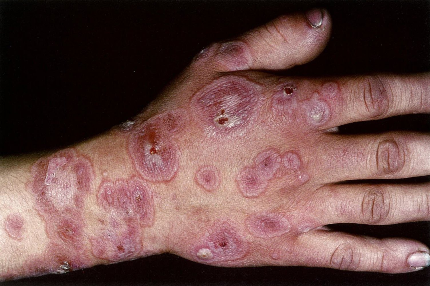
Multiple well-defined lesions can be seen on the back of the hand. The lesions consist of concentric rings with an outer erythematous zone, a paler intermediate zone, and a darker violaceous center, consistent with target lesions. Some of the lesions show central blistering.
This presentation is typical of erythema multiforme.
Source: © IMPP
Lower thigh and knee of a patient after antibiotic intake
Multiple erythematous, well-demarcated lesions can be seen. The lesions vary in size and shape, and some have merged to form larger lesions. The lesions show central erythema surrounded by a pale pink ring, characteristic of target lesions.
Source: © IMPP
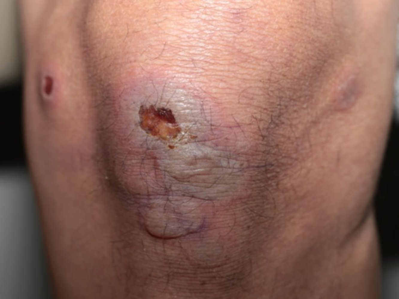
Multiple blistering lesions can be seen on the kneecap and sides of the leg. The lesions have a central ulcer surrounded by erythema, characteristic of blistering target lesions.
Source: Courtesy of Dr. Gary M. White, MD
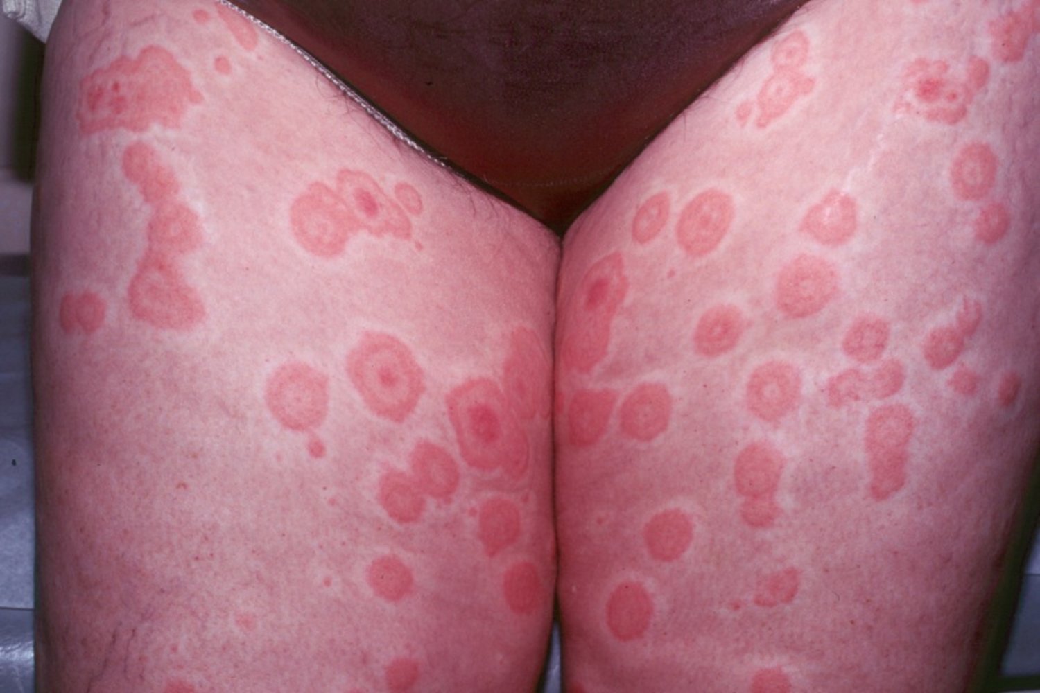
Multiple circular lesions with a central papule and raised edges (also called “target lesions”), surrounded by a lighter edematous area, are visible on the anterior aspect of the thighs.
This is the typical appearance of erythema multiforme.
Source: Courtesy of Dr. Gary M. White, MD
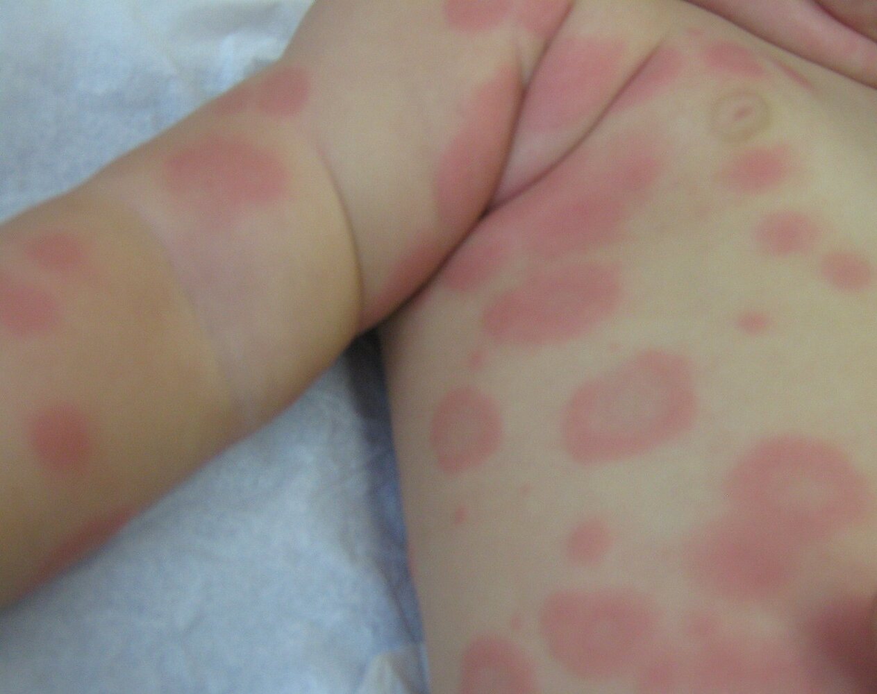
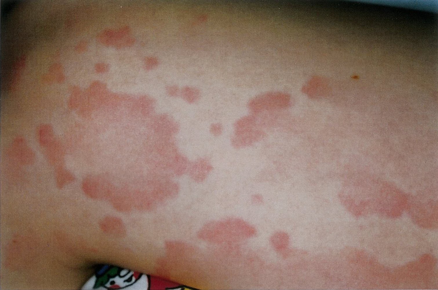
Trunk and right arm of an infant
Multiple erythematous macules are visible, arranged in concentric rings with a pale center and an outer erythematous margin. The macules are partly confluent and of different sizes.
These lesions are suggestive of erythema multiforme, a hypersensitivity reaction that can be caused by various infections (most commonly herpes simplex virus infections), certain drugs, or immune dysregulation.
Source: “Erythema multiforme EM 02” by Grook Da Oger, Wikimedia Commons, licensed under CC BY-SA 3.0.

Diagnosis
Erythema multiforme is primarily a clinical diagnosis based on the presence of typical lesions (e.g., target lesions) and patient history (e.g., exposure, recent infection). [4]
Laboratory studies [6]
-
Routine studies: Obtain in patients with erythema multiforme major; the following findings may be seen.
- Inflammatory markers (e.g., ↑ WBC, ↑ ESR)
- ↑ Liver enzymes
- Electrolyte abnormalities (e.g., due to insufficient intake of liquids and/or food)
-
Infectious disease workup: based on clinical suspicion
- Diagnosis of herpes simplex infection
- M. pneumoniae serology or PCR
- COVID-19 testing
-
Additional studies: Consider if there is diagnostic uncertainty to determine underlying causes and exclude other conditions. [4][6]
- Autoimmune studies, e.g.:
- Antinuclear antibody in suspected lupus erythematosus (Rowell syndrome)
- Serum complements in persistent erythema multiforme
- Malignancy workup (e.g., leukemia, lymphoma, solid organ neoplasms): especially in patients with recurrent erythema multiforme [6]
- Autoimmune studies, e.g.:
Skin biopsy [6]
- Indication: Consider if there is diagnostic uncertainty.
-
Supportive findings (nonspecific)
- Evidence of inflammation (e.g., lymphohistiocytic infiltrate)
- Necrotic keratinocytes
- Edema
-
Specific testing: Consider based on clinical suspicion.
- Autoimmune blistering diseases: direct fluorescent antibody test
- HSV infections: Tzanck smear test or PCR
Differential diagnoses
- Stevens-Johnson syndrome: In comparison to erythema multiforme, skin involvement in SJS is more severe and usually triggered by medications.
- Urticaria: The lesions of urticaria are transient, disappearing within a few hours, while those of erythema multiforme last up to a month; target lesions are uncommon.
- Prodrome phase of bullous pemphigoid: Prodromal rash resembles that of urticaria/erythema multiforme; oral involvement is rare.
-
Fixed drug eruption
- Definition: hypersensitivity reaction to specific drugs occurring within 8 hours of exposure
- Clinical features
- Well-defined, erythematous, pruritic lesions (often solitary)
- Oral mucosa may be affected; atypical target lesions may be present
- Resolves spontaneously on stopping the offending drug; may cause postinflammatory hyperpigmentation
- Diagnosis: provocation tests (oral challenge test/skin patch test)
- Rowell syndrome
-
See also:
- “Overview of annular skin lesions”
- “Maculopapular rashes”
- “Diffuse erythematous rashes”


References: [4][7][8]
The differential diagnoses listed here are not exhaustive.
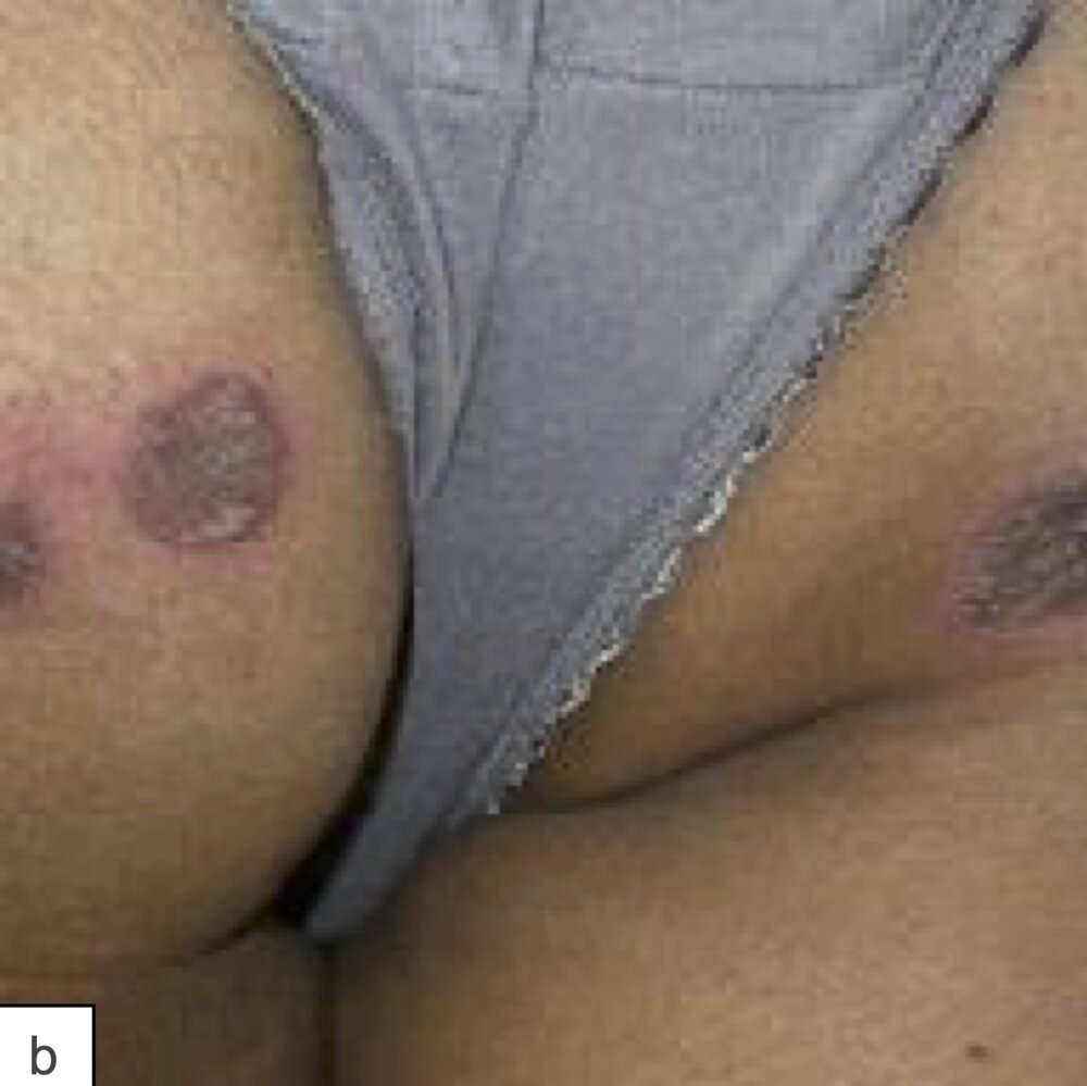
Well-defined, annular lesions on the left and right buttock with an erythematous rim around a hyperpigmented center are visible.
This characteristic presentation suggests a fixed drug eruption with possible post-inflammatory hyperpigmentation. Fixed drug eruptions are characterized by a recurrence at the same site when reexposure to the offending substance occurs.
Source: “Figure 94 b: Fixed drug eruption; in: Atlas of Pediatric HIV Infection” by Regina E. Oladokun; Rannakoe J. Lehloenya; Carol Hlela et al., Openbooks, licensed under CC BY-ND 4.0.
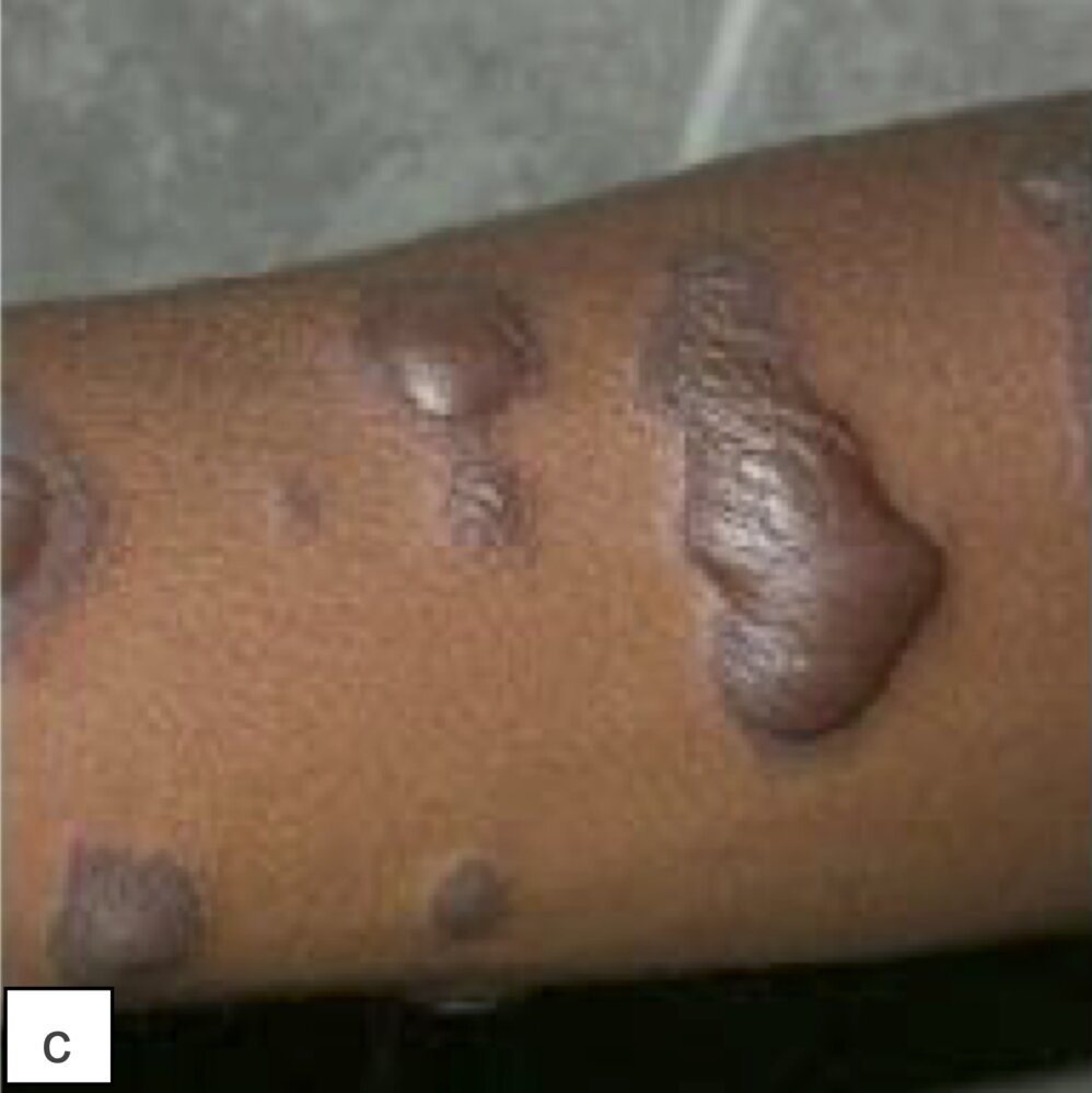
Multiple, heterogeneous, and mostly bullous lesions with characteristic, slight erythematous rims around a hyperpigmented center are visible.
While blisters and/or ulcerations of the damaged skin may not always occur, the rest of the highlighted findings are still suspicious for a fixed drug eruption.
Source: “Figure 94 c: Classic round pigmented {...}; in: Atlas of Pediatric HIV Infection” by Regina E. Oladokun; Rannakoe J. Lehloenya; Carol Hlela et al., Openbooks, licensed under CC BY-ND 4.0.
Treatment
General principles [4][6]
- Treatment is primarily symptomatic.
- Discontinue any offending medications.
- Consider treating any underlying infection (e.g., treatment of HSV infection, antibiotic therapy for M. pneumonie).
- Consider systemic glucocorticoid therapy.
-
For patients with erythema multiforme major with severe mucosal involvement, consider:
- IV fluid therapy and electrolyte repletion (management is similar to that of extensive burns)
- Specialized nutrition support
- Antibiotic therapy for SSTIs
- Specialist consultation (e.g., ICU, dermatology, ophthalmology)
Patients with erythema multiforme major and severe oral mucosal involvement may suffer from dehydration and/or electrolyte abnormalities due to insufficient intake of liquids and food. [6]
Most patients with erythema multiforme minor require no specific treatment because the condition is self-limited. [4]
Pharmacotherapy [2][4][6]
Antiviral therapy
- Acute erythema multiforme episode: Consider antiviral treatment of HSV infection (uncertain benefit). [2][4][6]
- Recurrent erythema multiforme: Long-term antiviral suppression therapy (e.g., acyclovir (off-label) for 6 months) is recommended as prophylaxis.
Pharmacotherapy (e.g., antivirals, systemic glucocorticoids) is recommended in patients with recurrent erythema multiforme to prevent further episodes. [4][6]
Supportive care [2]
-
Systemic glucocorticoids
- Consider in patients with erythema multiforme major with severe mucosal involvement. [2][4][6]
- Example agent: prednisone with a prolonged taper over 2–4 weeks [2][6]
- Topical glucocorticoids (e.g., hydrocortisone )
- Antihistamines for pruritus (e.g., hydroxyzine )
-
Mucosal care, e.g.: [2]
- Oral anesthetics, antiseptic solution, and/or high-potency hydrocortisone gel
- Ophthalmology consultation and ophthalmic medications (e.g., topical lubricants)
- Pain management: See “Oral analgesics” and “Parenteral analgesics” for dosages.
Prognosis
- Erythema multiforme is usually self-limited and occurs as a single episode; rash spontaneously disappears within 1 month. [4]
- Some patients may develop either: [4]
- Recurrent erythema multiforme
- Persistent erythema multiforme
References
- Lamoreux MR, Sternbach MR, Hsu WT. "Erythema multiforme". Am Fam Physician. 74(11). :1883-1888. (2006)
- Soares A, Sokumbi O. "Recent Updates in the Treatment of Erythema Multiforme". Medicina. 57(9). :921. (2021)
- Kaur S, Handa S. "Erythema multiforme following vaccination in an infant". Indian J Dermatol Venereol Leprol. 74(3). :251-253. (2008)
- Trayes KP, Love G, Studdiford JS. "Erythema Multiforme: Recognition and Management". Am Fam Physician. 100(2). :82-88. (2019)
- Lerch M, Mainetti C, Terziroli Beretta-Piccoli B, Harr T. "Current Perspectives on Erythema Multiforme". Clin Rev Allergy Immunol. 54(1). :177-184. (2018)
- Sokumbi O, Wetter DA. "Clinical features, diagnosis, and treatment of erythema multiforme: a review for the practicing dermatologist". Int J Dermatol. 51(8). :889-902. (2012)
- Valente Duarte de Sousa IC. "Fixed drug eruption". N Engl J Med. 365. :e12. (2011)
- "Fixed Drug Eruption". http://www.dermnetnz.org/topics/fixed-drug-eruption/. [2001-01-01]